OUR WORK
|
|
|
|
|
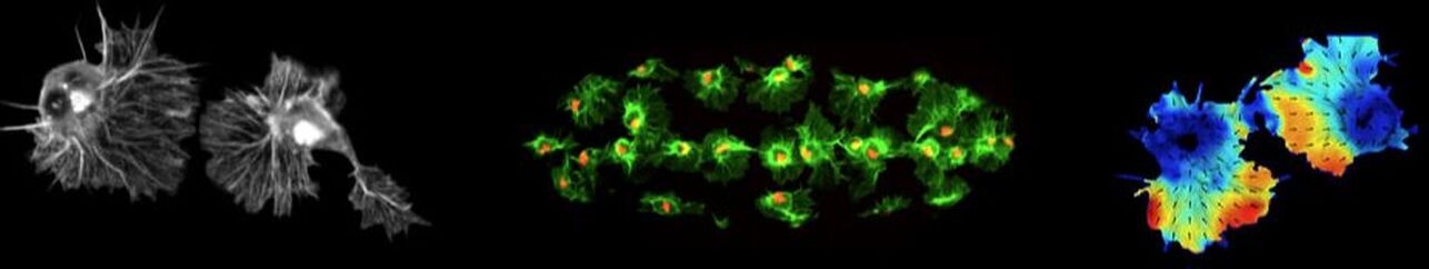
One of the interests of the laboratory is to understand how hemocytes maintain their even spacing within the embryo. We have shown that during their migration, hemocytes are undergoing contact inhibition of locomotion. This process of collision and subsequent repulsion is an instructive migratory cue that allows the cells to maintain space within the embryo. We are now investigating the cytoskeletal machinery and cellular recognition mechanisms that allow hemocytes to undergo this process with such reproducibility. This movie shows an example collision between two colliding hemocytes in which their actin and microtubule cytoskeletal networks are labeled.
|
|
|
Hemocytes can be imaged within the living Drosophila embryo at a resolution approaching what can be obtained from cells in culture. This has opened up the possibility of more detailed image analysis techniques that allow for subcellular quantification of protein dynamics within these cells. This movie shows colliding hemocytes in which their actin cytoskeleton was fluorescently labelled. Subsequently, the dynamics of the flowing actin networks was quantified by particle image velocimetry, which allows us to accurately measure the direction and speed of actin motion within these cells. Such computer vision techniques will allow us to precisely determine the intracellular mechanisms driving hemocyte movement.
|
|
|
|
Work in the laboratory is also revealing the function of hemocyte migration for Drosophila development. Recent work has shown that the reason for hemocyte dispersal is to allow these cells to evenly distribute extracellular matrix in the embryo. This movie shows embryonic hemocytes depositing Collagen IV, one of the major extracellular matrix components of the basement membrane. This is a surprising function for these cells, as extracellular matrix production is not thought to be a major function for macrophage populations. We are now using this model to understand how the basement membrane is constructed de novo during development and examine the function of this extracellular matrix for tissue morphogenesis.
|
Stramer Lab |
King’s College London
Randall Centre for Cell & Molecular Biophysics New Hunt's House, Guy's Campus London SE1 1UL United Kingdom |
Professor Brian Stramer
|